Minimally Invasive Spinal Fusion: Thoracic and Lumbar

HeaderText
What is Minimally Invasive Spinal Fusion?

Minimally = as little as possible
Invasive = intrusive

Spinal = having to do with the spine
Fusion = a process in which bones “fuse,” or grow together into one bone

Thoracic = having to do with the spine in the upper and middle back
Lumbar = having to do with the spine in the lower back

Spinal fusion is a procedure in which grafted (transplanted) bone grows together with bone already in the spinal column, forming one solid bone and stabilizing that section of spine. When performed with a minimally invasive approach, the procedure uses specialized instruments and techniques that minimize cutting and other disruption of the body’s tissues.

Fusion of the lumbar spine means spinal fusion in the lower back. Fusion of the thoracic spine means spinal fusion in the upper or middle back. Fusion is more common in the lumbar than the thoracic spine

When is this Procedure Performed?

Thoracic and lumbar fusion are performed to restore strength and stability to those sections of spine. This may be necessary for an adult or pediatric patient when deformity, degeneration, tumor, or trauma renders the spine unstable. In such cases, a neurosurgeon will realign the spine if necessary, then perform a fusion.

In other cases, another medical intervention may have the unwanted side effect of making the spine unstable. Instability as a result of medical intervention is called iatrogenic instability. For example, the surgical treatment of a tumor, a degenerated disc, or a deformity may require the removal of certain sections of spinal disc or bone. This surgery treats the presenting problem, stopping the pain or other neurological symptoms, but leaves the spine less stable. A neurosurgeon typically performs a fusion during the same operation that would otherwise produce iatrogenic instability.

How is this Procedure Performed?

Minimally invasive thoracic and lumbar fusion can be accomplished with the following procedures:

  • Transforaminal interbody lumbar fusion (TLIF) – using this minimally invasive technique, the surgeon can achieve fusion of both the front and back parts of the spine during one procedure.
  • Lateral lumbar interbody fusion (LLIF) (also known as XLIF tm) – using this minimally invasive technique, the surgeon approaches the spine from the side. This approach produces the least disruption to muscles, bones, and abdominal organs.

In adults and children, minimally invasive spinal fusion is performed under general anesthesia, which means the patient is unconscious.

In minimally invasive surgery, the surgeon makes small incisions. The number, location, size and shape of the incisions vary depending on the location of the problem and the approach the surgeon has selected.

For most types of minimally invasive spinal fusion, the surgeon uses instruments called tubular dilators. These are tubes of expanding diameter that move aside the muscles and other tissues located between the skin incision and the spine. The dilators create a tunnel through these tissues down to the spinal column. An instrument called a tubular retractor holds the tunnel open while the surgeon works. Some surgical procedures require more than one retractor.

Unlike in some traditional surgical procedures, in minimally invasive procedures, surgeons may have a limited view of the surgical area. In order to see the area, surgeons may use an operating microscope positioned at the top of the retractor, or an endoscope (thin tube with a camera and a light at the end) that passes down through the retractor into the body. The surgeries also tend to rely more upon intraoperative radiographic imaging to guide the surgeon.

If another minimally invasive procedure must be performed during the same operation, it is performed first. This is often a decompression procedure, performed to relieve pressure on the spinal cord or surrounding nerves. Bone or disc sections removed during a minimally invasive decompression are extracted through the tubular retractors.

Then the surgeon performs the spinal fusion through the retractor. To fuse the vertebrae, the surgeon places new bone material, called bone graft, as a bridge between existing bones. The graft may come from a bone bank or it may be taken from the patient’s own body.  Sometimes bone removed from the patient during the decompression portion of the surgery can be used as a graft; other times, the bone is taken from the patient’s iliac crest (hip bone). Depending on the type of surgery, bone from the hip may be removed through the same surgical incision or through a separate incision.

To aid in bony fusion, the surgeon may also use a “bone promoting substance” like bone morphogenic protein, or BMP. BMP aids in bone growth and is naturally produced by the body. Other materials, like synthetic bone graft extenders or processed bone (called demineralized bone matrix) may also be used in combination with the bone graft and/or BMP. These materials can add volume to the bone graft without the need to harvest more bone. Newer technology involves using concentrated bone cells or stem cells either harvested from the patient or from a donor. The use of some of these substances is controversial in pediatric patients; speak with a pediatric neurosurgeon for the most up-to-date information and recommendations for particular cases.

Most surgeons also insert hardware such as screws, rods or plates as part of a fusion procedure in order to hold the bones in place until they heal. This procedure is called fixation. Fixation is also performed through the retractor. The surgeon then removes the retractor and closes the incision.

In some cases, a back brace will be prescribed to hold the spine in one position while the bones begin to fuse.

How Should I Prepare for this Procedure?

Nicotine reduces the body’s ability to create fusion between the bones. If you smoke or use tobacco products, talk to your neurosurgeon about quitting before your surgery. Using tobacco products increases the probability of a failed fusion.

Make sure to tell your doctor about any medications or supplements that you’re taking, especially medications that can thin your blood such as aspirin. Your doctor may recommend you stop taking these medications before your procedure. To make it easier, write all of your medications down before the day of surgery.

Be sure to tell your doctor if you have an allergy to any medications, food, or latex (some surgical gloves are made of latex).

On the day of surgery, remove any nail polish or acrylic nails, do not wear makeup and remove all jewelry. If staying overnight, bring items that may be needed, such as a toothbrush, toothpaste, and dentures. You will be given an ID bracelet. It will include your name, birthdate, and surgeon’s name.

What Should I Expect After the Procedure?

How long will I stay in the hospital?
Patients are encouraged to walk on the day following surgery, and typically stay in the hospital 1-3 days.

Will I need to take any special medications?
You will be prescribed pain medications to help manage post-surgical pain.

Will I need to wear a brace?
A brace may be worn for comfort after surgery, but is not required in most patients.

When can I resume exercise?
In the first stages of healing, 1-2 months after surgery, there is no advantage to pushing the limits in terms of physical activity. Short periodic walks are sufficient to prevent the medical complications associated with inactivity. The most important thing you can do in the beginning is be patient and allow your body time to heal.

After this initial healing period, you will undergo a gradual return to activity guided by your doctor.

What follow-up will I receive?
The surgeon will schedule the first follow-up visit 4-6 weeks after surgery, and periodically thereafter. X-rays are typically performed during the follow-up period to monitor how the bones are fusing.

Will I need rehabilitation or physical therapy?
Physical therapy is often useful. It usually begins several weeks after surgery and focuses on lower back strengthening and increasing range of motion.

Will I have any long-term limitations due to thoracic or lumbar spinal fusion?
There may be some reduction in the range of motion and mobility of the fused spinal segments. This is minimized by an active exercise and stretch program followed by the healing of the spinal fusion.

After your fusion surgery, it is essential that you continue a healthy spine care lifestyle, including achieving and maintaining your ideal weight, implementing an active core strengthening program, and abstaining from all tobacco products.


Preparing for Your Appointment

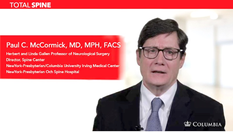
Drs. Paul C. McCormick, Michael G. Kaiser, Peter D. Angevine, Alfred T. Ogden, Christopher E. Mandigo, Patrick C. Reid and Richard C. E. Anderson (Pediatric) are experts in minimally invasive spinal fusion.


Helpful Surgery Overviews

Dr. McCormick will choose the treatment method specific to each patient and situation. Some of the condition’s treatment options may be listed below.

Recent News