| Header | Text |
| Summary | Arachnoid = one of the protective membranes around the brain and spinal cord
Cyst = a collection of fluid
Arachnoid cysts are fluid-filled sacs that may develop in a membrane around the brain or spinal cord. At the Spine Hospital at the Neurological Institute of New York, we specialize in spinal arachnoid cysts. (For more information about arachnoid cysts in the brain, click here.)
The spinal cord and brain are bathed and protected by a fluid called cerebrospinal fluid (CSF). Three layers of membrane cover the brain and spinal cord, sealing in the CSF. When the CSF builds up inside one of the layers, called the arachnoid, the result is an arachnoid cyst.
Arachnoid cysts can be found at any level of the spine: the cervical spine (spine in the neck), the thoracic spine (in the upper and mid back), and lumbar spine (in the lower back).
|
| Symptoms | Symptoms of a spinal arachnoid cyst depend on its size and location. Many spinal arachnoid cysts cause no symptoms at all. However, if the cyst is large enough, it can compress the spinal cord and surrounding nerves. In these cases, patients may experience pain, weakness, or numbness in the back, arms, or legs.
Large arachnoid cysts in the lumbar spine may also cause loss of bladder and/or bowel control (cauda equina syndrome), cramping or pain with walking (neurogenic claudication), or sharp pain that radiates through the buttock, hip, leg and/or foot (sciatica).
If left untreated, spinal arachnoid cysts may cause permanent severe neurological damage.
|
| Causes and Risk Factors | Several theories exist about the exact cause(s) of spinal arachnoid cysts and the mechanism(s) of their development. Research is still ongoing, and to date no one is certain which theory is correct.
Researchers do know that most spinal arachnoid cysts begin during infancy. They are the result of a congenital (in-born) problem with the arachnoid layer. Referred to as primary cysts, these are usually found in the thoracic spine (upper and mid back).
However, some spinal arachnoid cysts, called secondary cysts, develop later in life. These cysts develop after an infection, trauma, bleeding, or surgery. They are more common in males than in females, and can appear at any spinal level.
|
| Tests and Diagnosis | Spinal arachnoid cysts are often found by chance when patients undergo testing for other conditions. This is called an incidental finding. Most spinal arachnoid cysts found incidentally cause no symptoms and need no treatment.
However, if a spinal arachnoid cyst has not been identified and a patient presents with symptoms associated with the condition, the doctor may order the following diagnostic procedures:
- Computed tomography myelography (myelo-CT) – uses opaque dye, X-rays and a computer to provide a detailed image of the spinal cord and nerve roots
- Magnetic resonance (MR) imaging – provides detailed images of the spine and spinal canal. MR imaging scans are very useful, because they can help a doctor determine whether a spinal arachnoid cyst interferes with the spinal cord and surrounding nerves.
Note that the symptoms of a spinal arachnoid cyst are similar to symptoms of other conditions that interfere with the spinal cord or nearby nerves. Before arriving at a definitive diagnosis, a doctor may need to rule out other problems, such as tumor, infection, or herniated disc. At the same time, spinal arachnoid cysts may be present without causing any symptoms at all. If a cyst is discovered, but it does not explain a patient’s symptoms, the cyst may not be the source of symptoms.
|
| Treatments | When a spinal arachnoid cyst causes symptoms, surgery may be necessary to remove it.
Some types of cysts can be removed completely, and the defect that allowed them to form can be closed.
In other cases, this approach is not possible. Then the neurosurgeon may perform one of the following procedures:
- Shunt Placement: This surgical procedure involves the placement of a permanent drainage system (cystoperitoneal shunt) to remove pressure from the cyst.
- Cyst fenestration: This surgical procedure involves opening the cyst and letting the fluid drain out.
Choice of surgical procedure depends on factors such as the cyst type, size, and location. An experienced neurosurgeon can determine the best procedure for each patient.
|
| Preparing for Your Appointment | At The Spine Hospital at The Neurological Institute of New York our spinal neurosurgeons are experts in the evaluation and management of patients with an arachnoid cyst.
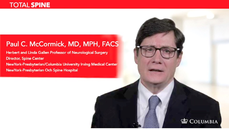
Drs. Paul C. McCormick, Michael G. Kaiser, Peter D. Angevine, Alfred T. Ogden, Christopher E. Mandigo, Patrick C. Reid, Richard C.E. Anderson (Pediatric), and Neil A. Feldstein (Pediatric) are experts in treating arachnoid cysts. They can also offer a second opinion.
|


