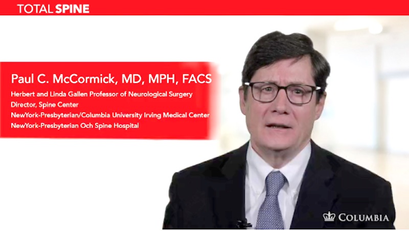
In his latest installment to the Total Spine series of instructional videos, Dr. Paul McCormick describes retropleural thoracotomy, a surgical technique used to treat complex spinal conditions of the thoracic and lumbar spine. “Retropleural thoracotomy is an important...
Ependymoma
Header | Text |
Summary | An ependymoma is a tumor that arises from ependymal cells, support cells in the brain and spinal cord. At the Spine Hospital at the Neurological Institute of New York, we specialize in spinal ependymomas. Ependymomas are intramedullary tumors, which means they arise within the substance of the spinal cord itself. They may occur at any level of the spinal cord, including the cervical (neck), thoracic (upper back), and lumbar (lower back). Some ependymomas, known as myxopapillary filum terminale ependymomas, arise in the lower lumbar and sacral region (base of the spine, above the tailbone) in the filum terminale. This structure is is a delicate, fibrous strand that extends below the spinal cord and helps support it. Ependymomas in this location can be difficult to remove safely. |
Symptoms | Spinal ependymomas arise from ependymal cells located in the very center of the spinal cord. They slowly expand over a prolonged period, often years, gradually putting pressure on the surrounding spinal cord. This compression of the spinal cord may cause symptoms of pain at the tumor site; numbness or pins and needles in the arms or legs; or weakness or clumsiness in the arms or legs. Exact symptoms depend on the tumor’s size and location. Symptoms vary considerably in terms of onset and progression. They may be minimal, intermittent or non-progressive for prolonged periods. The benign and slow-growing nature of intramedullary spinal cord tumors means symptoms may be absent for a long time while the tumor is smaller. This may delay patient presentation for medical assessment and diagnosis. Fortunately, most patients are now initially diagnosed with little or no neurological abnormality. Exceptions exist, and sometimes the onset and progression of symptoms can occur over a shorter period of time. |
Causes and Risk Factors | The causes of ependymoma are not yet understood. Men and women are equally affected, and most ependymomas occur in the 4th through 6th decades of life. But the tumors can develop in people of all ages, races, and sexes. People with a genetic condition called neurofibromatosis type 2 are predisposed to developing ependymomas. |
Tests and Diagnosis | The diagnosis of ependymoma is usually made with magnetic resonance (MR) imaging. MR scans use a combination of large magnets, radio waves, and a computer to produce detailed images of organs and structures inside the body. They are sometimes performed with intravenous contrast enhancement. In an MR with intravenous contrast enhancement, a special tracer–a substance like a dye that is visible to the MR scan–is injected into the blood vessels. MR scans with contrast enhancement reveal an ependymoma as a well- defined nodule within the spinal cord. Cysts that extend either below or above the tumor are common, and spinal cord expansion or swelling may also be seen. |
Treatments | Due to the benign, slow-growing nature of intramedullary tumors, the mere presence of an ependymoma is not necessarily an indication for immediate treatment. Often these tumors are discovered incidentally (in passing) in patients without any symptoms. If the tumor is small and does not exert pressure on the surrounding spinal cord, it may simply be followed with annual MR imaging. Several years may pass before the tumor begins to put enough pressure on the spinal cord to necessitate treatment. For larger tumors, or those tumors causing symptoms, treatment is recommended. Because these tumors are benign and slow-growing, microsurgical tumor removal is the treatment of choice for most patients. In microsurgical tumor removal, a surgeon uses a surgical microscope and very fine instruments to expose and remove the tumor. Microsurgery to remove an ependymoma is performed under general anesthesia with the patient positioned face-down. Spinal cord function is carefully monitored throughout the procedure using precise electrophysiological techniques such as SSEP (somatosensory evoked potentials) and MEP (motor evoked potentials). A laminectomy, or removal of a portion of the back of the spine, is performed to gain access to the spinal canal. The thin covering of the spinal canal known as the spinal dura is opened to expose the spinal cord. A thin opening in the back portion of the spinal cord is made to expose the tumor. Ependymomas are usually well defined and can be separated from the surrounding spinal cord using neurosurgical techniques. A laser or ultrasonic tool may be utilized. In most patients, the tumor can be completely removed without substantially disturbing the function of the spinal cord. However, many patients may note some diminished sensation in the legs due to the opening of the spinal cord. A more complete description and illustration of the surgical techniques are demonstrated in this video. Watch Dr. McCormick perform the surgery in this video.
|
Preparing for Your Appointment | Drs. Paul C. McCormick, Michael G. Kaiser, Peter D. Angevine, Alfred T. Ogden, Christopher E. Mandigo, Patrick C. Reid, Richard C.E. Anderson (Pediatric), and Neil A. Feldstein (Pediatric) are experts in treating ependymomas. They can also offer you a second opinion. |
Helpful Surgery Overviews
Dr. McCormick will choose the treatment method specific to each patient and situation. Some of the condition’s treatment options may be listed below.
Recent News
Columbia Neurosurgeons Named New York’s Super Doctor
https://www.neurosurgery.columbia.edu/news/columbia-neurosurgeons-named-new-yorks-super-doctor
Dr McCormick video on microsurgical resection of a synovial cyst causing severe lumbar spinal stenosis published in Operative Neurosurgery
Synovial Cysts are a benign and fairly common condition in adult patients. They can arise in most joints throughout the body. In most cases they are asymptomatic but in the spine they can enlarge to produce severe spinal stenosis and pressure on the spinal nerves...
Dr. McCormick invited speaker at 37th Annual Meeting of the Joint Section on Disorders of the Spine and Peripheral Nerves
Dr. McCormick was a featured speaker at the recent 37th Annual Joint Spine Section Annual Spine Summit meeting held in San Diego. He presented at the Innovative Technology Special Session: Operative Video Segments. His presentation was entitled “Intramedullary Tumor,...