Chiari Malformation

HeaderText
Summary

A Chiari malformation is a congenital (present at birth) defect in the area of the back of the head where the brain and spinal cord connect. The condition is also called Arnold Chiari malformation. There are four types of Chiari malformations, including the following:

Type I – this is the most common type of Chiari malformation. Commonly goes unnoticed until problems arise in the adolescent or adult years of life. In this condition, the base of the skull and the upper spinal area are not formed properly.

Type II – In this condition, part of the back of the brain shifts downward through the bottom of the skull area.

  • Type II Chiari malformations are typically seen in infants who are born with spina bifida, a neurological condition that causes a portion of the spinal cord and the surrounding structures to develop outside, instead of inside, the body.
  • Type II Chiari malformations can also be associated with a condition known as hydrocephalus. Hydrocephalus is a condition in which there is an overproduction or lack of absorption of the cerebral spinal fluid (CSF) that is found inside of the ventricles (fluid-filled areas) inside of the brain. The increased fluid causes the pressure inside of the head to increase and the skull bones to expand to a larger-than-normal appearance.

Type III – The back of the brain protrudes out of an opening in the back of the skull area.

Type IV – The back of the brain fails to develop normally.

Symptoms

The following are the most common symptoms of a Chiari malformation. However, each child may experience symptoms differently. In infants and older children born with this condition, symptoms may include:

  • Headaches
  • Stiffness or pain in the neck or back of the head area
  • Poor feeding and swallowing
  • Decreased strength in the arms
  • Decreased sensation in the arms and legs
  • Rapid, back and forth, eye movement
  • Developmental delays
  • Weak cry
  • Breathing problems

The symptoms of Chiari malformation may resemble other conditions or medical problems. Always consult your child’s physician for a diagnosis.

Causes and Risk Factors

Although the exact cause of Chiari malformation is unknown, it is thought that a problem during fetal development may cause the abnormal brain formation. Chiari malformation may be caused by exposure to harmful substances during fetal development or associated with genetic problems or syndromes that may have a tendency to run in families.

Theories suggest that the following may predispose the fetus to problems that affect the normal development of the head during pregnancy:

  • Exposure to hazardous chemicals/substances
  • Lack of proper vitamins and nutrients in the diet
  • Infection
  • Prescription or illegal drug and alcohol consumption
Tests and Diagnosis

If a Chiari malformation occurs with other congenital (present at birth) defects, the diagnosis may be made at birth. Other times, the diagnosis is made after the onset of specific signs and symptoms, and after diagnostic testing. The physician obtains a complete prenatal and birth history of the child and may also ask if there is a family history of any medical problems. The physician will also ask about developmental milestones, such as the age the child sat up, crawled, or walked since a Chiari malformation can be associated with other neuromuscular disorders. Developmental delays may require further medical follow up for underlying problems.

During the examination, a measurement of the circumference of the child’s head is taken and compared to a scale that can identify normal and abnormal ranges.

Diagnostic tests that may be performed to confirm the diagnosis of a Chiari malformation include:

  • Magnetic Resonance Imaging (MRI) – a diagnostic procedure that uses a combination of large magnets, radiofrequencies, and a computer to produce detailed images of organs and structures within the body.
Treatments

There are many ways to treat Chiari malformations, but all require surgery. The basic operation is one of uncrowding the area at the base of the cerebellum where it is pushing against the brainstem and spinal cord. This is done by removing a small portion of bone at the base of the skull deep to the neck muscles as well as often removing a part of the back of the first and occasionally additional spinal column segments. The operation is often modified if there is a syrinx present or if the child has hydrocephalus. Most children who have the surgery do quite well and have  improvement of their symptoms.

At the Children’s Hospital of New York, we have been collecting information since 1998 using intraoperative electrophysiological monitoring to help determine whether opening of the dura (a thick membrane that surrounds the brain and spinal cord) is a necessary component of surgery for children with Chiari I malformation. We discovered that most of the improvement in nerve impulses through the brain and spinal cord occurs after removal of the bone. However, we did not see any further improvement after opening the dura, suggesting that children may not require this additional step of surgery. Accordingly, for the past four years we have performed a less invasive operation where the dura is not opened during surgery. At this time, we have seen excellent clinical and radiographic results without any significant operative complications after bony decompression without dural opening. This is important because the complication rate after surgery has been reported to be nearly 4 times higher if the dura is opened during surgery.

In addition, the use of endoscopes has allowed for this procedure to be performed through smaller incisions, which helps in the reduction of post operative pain and speech recovery.

Specific treatment for a Chiari malformation will be determined by your child’s physician based on:

  • Your child’s age, overall health, and medical history
  • The extent of the condition
  • The type of condition
  • Your child’s tolerance for specific medications, procedures, or therapies
  • Expectations for the course of the condition
  • Your opinion or preference

Medical management consists of frequent physical examinations and diagnostic testing to monitor the growth and development of the brain, spinal cord, skull, and backbones.

Some types of Chiari malformations may require surgery to relieve increased pressure inside the head or neck area, or to help drain excess cerebral spinal fluid from the brain. Very severe Chiari malformations may be life threatening.

Parents are instructed to watch for any changes that may affect the child’s neurological status, including the following:

  • Breathing problems
  • Degree of alertness
  • Speech or feeding problems
  • Problems walking
  • Uncoordinated movement

Life-long considerations for a child with a Chiari malformation:

The full extent of the problems associated with a Chiari malformation are usually not completely understood immediately at birth, but may be revealed as the child grows and develops. Children born with a Chiari malformation require frequent examinations and diagnostic testing by his/her physician to monitor the development of the head as the child grows. The medical team works hard with the child’s family to provide education and guidance to improve the health and well-being of the child.

Genetic counseling may be recommended by the physician to provide information on the recurrences for Chiari malformation and any available testing.

Preparing for Your Appointment

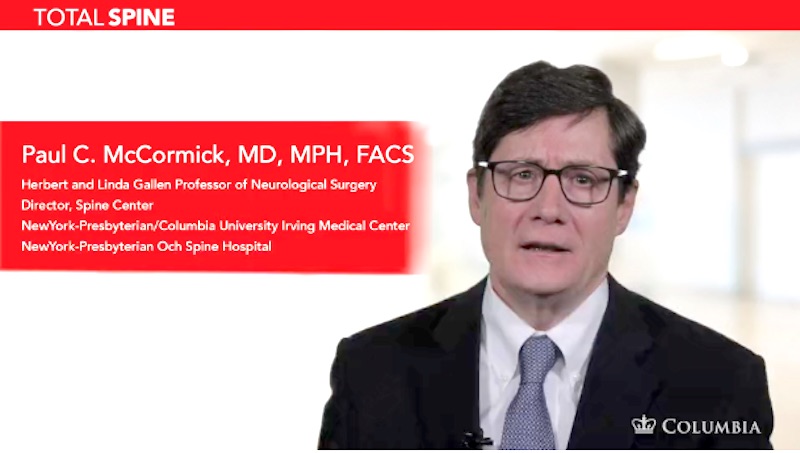
Drs. Paul C. McCormick, Patrick C. Reid, Richard C. E. Anderson (Pediatric), and Neil A. Feldstein (Adult & Pediatric)  are experts in treating chiari malformations. They can offer you a second opinion.

Helpful Surgery Overviews

Dr. McCormick will choose the treatment method specific to each patient and situation. Some of the condition’s treatment options may be listed below.

Recent News